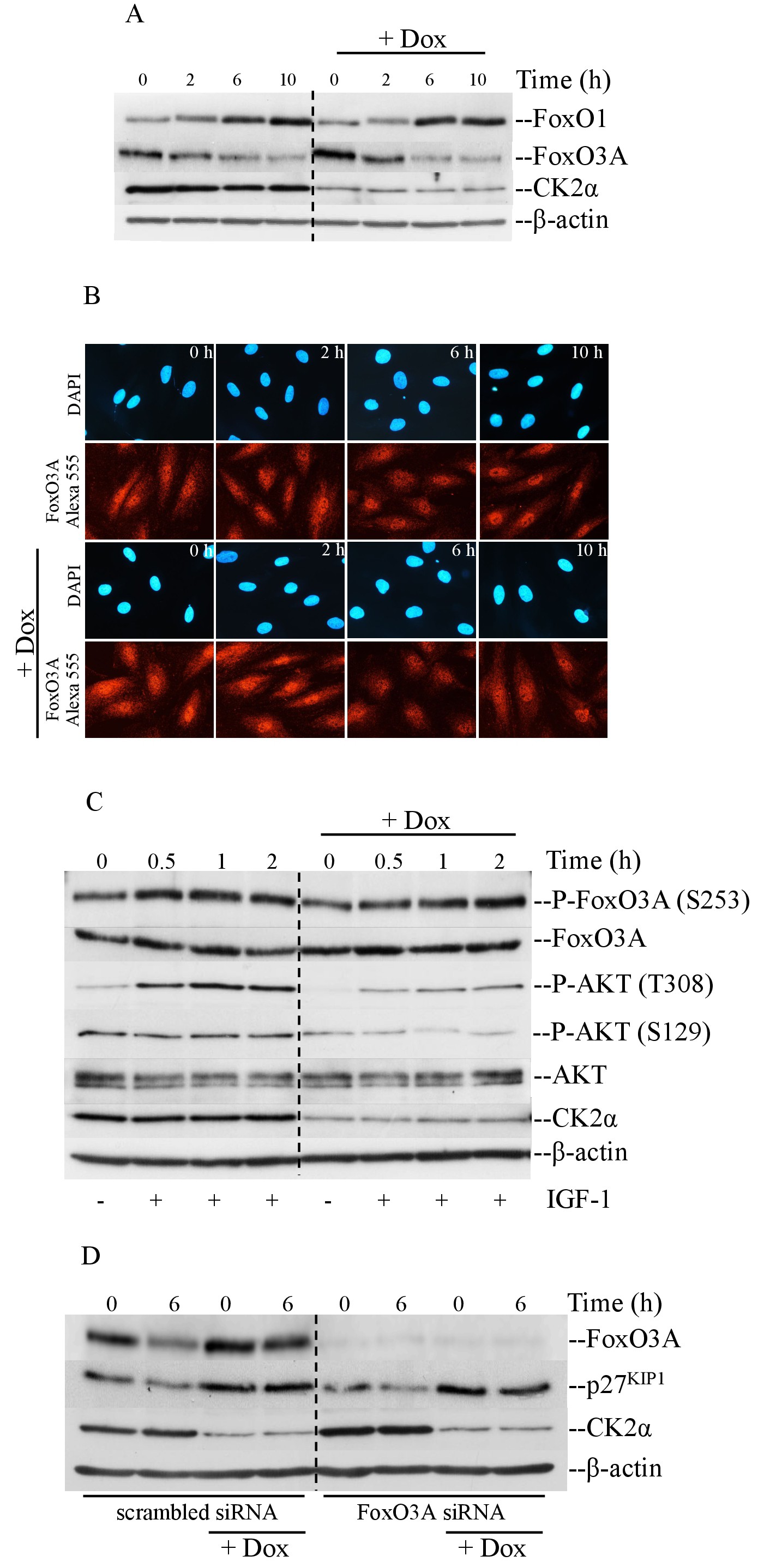
Fig. 6. Up-regulation of p27KIP1 correlates with slightly higher levels of FoxO3A in cells with lowered expression of CK2α. However, FoxO3A silencing does not affect the expression of p27KIP1. (A) Cells were serum-starved for 48 hours in the presence or absence of doxycycline and harvested at the indicated time points. Whole cell lysate was analyzed by western blot employing antibodies against FoxO1 and FoxO3A, respectively. Detection of β-actin confirmed equal protein loading. (B) Cells treated as indicated in (A) were labeled with rabbit polyclonal anti-FoxO3A. Proteins were visualized by staining with Alexa Fluor 555-conjugated streptavidin (red fluorescence) after incubation with a secondary anti-rabbit IgG antibody conjugated with biotin. Cellular DNA was detected by staining with DAPI reagent (blue fluorescence). Cell pictures were taken at 40x magnification. (C) Cells were rendered quiescent by serum withdrawal. Subsequently, they were stimulated by adding 100 ng/ml IGF-I and harvested at the indicated time points as shown in the Fig.. Whole cell extract was analyzed by western blot employing antibodies against the indicated proteins. (D) Down-regulation of FoxO3A was obtained by transfecting cells with siRNA targeting FoxO3A-mRNA. Control cells were transfected with scrambled siRNA. Cells were synchronized by serum starvation and harvested after 0 hours (i.e. corresponding to quiescent cells) and 6 hours after stimulation. Experiments were repeated at least three times obtaining similar results. One representative experiment is shown.